
14.02.2017 · 👁 5,795
MRI department of the clinic “SALUS VITA”
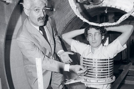
MRI was first introduced into clinical medicine more than 30 years ago. Since then, significant progress has been made both in technology and in clinical applications of the method, especially in the last 10 years. MRI has become a key imaging modality and is very effective in diagnosing a wide variety of diseases. In recent years, the methodology for examining the chest organs, including the heart, abdominal cavity, genitourinary and musculoskeletal systems, has been optimized. MR imaging scanners with higher magnetic field strengths and whole body MRI have become a clinical reality.
Due to this continuous dynamic development, of course, there is a need for continuous training. This method combines painlessness, safety for health and high accuracy of the data obtained. MRI is a non-invasive diagnostic method; it does not require direct intervention in the human body. Today MRI is used in many branches of medicine. There are various types of MRI diagnostics. Based on the part of the body being examined, MRI can be divided into:
MRI of the brain. Used in neurosurgery and neurology. Helps to accurately determine pathologies of brain tissue. Before the operation, it helps to specify the extent and specific location of the intervention.
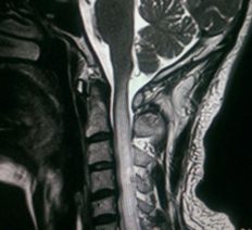
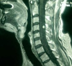
MRI of the spine (cervical, thoracic, lumbar and lumbosacral regions). Allows you to identify a wide range of diseases of the spinal cord and spinal region. Using this type of MRI, it is possible to calculate the location and size of a disc herniation, tumor or spinal injury with millimeter precision.
MRI of the abdominal cavity and retroperitoneum (liver, kidneys, adrenal glands, spleen, pancreas, etc.). Allows you to obtain information about the condition of all organs of the abdominal cavity and retroperitoneal space. Using MRI images, many pathologies of this group of organs can be diagnosed in the early stages.
MRI of the pelvic organs. Most often used in gynecology and urology. Well suited for diagnosing female diseases. In men, this type of MRI examines the prostate. In addition, for men, an MRI of the external genitalia is performed to examine the testicles.
MRI of joints (knee, hip, elbow, shoulder, etc.). Used in orthopedics, surgery and traumatology. Large joints are most often examined. MRI helps determine the condition of cartilage surfaces, joint capsules, ligaments, tendons, muscles, etc.
MRI angiography of vessels. Usually the vessels of the brain, upper and lower extremities, and internal organs are examined. MRI allows you to evaluate the functionality of the circulatory system. Both veins (venography) and arteries (arteriography) are examined.
Another type of magnetic resonance imaging worth mentioning is MRI with a contrast agent. Contrast material increases the diagnostic sensitivity of MRI. For some types of MRI diagnostics, a contrast agent is simply necessary (search for tumors, study of blood vessels, etc.). Paramagnets are used as contrast.
Thus, MRI is a reliable method for diagnosing various areas. Tomography is used in oncology, traumatology, surgery, orthopedics and gynecology to successfully resolve a variety of problems.
Doctors have one of the noblest professions because they take care of us when we are sick and think about our health with deep empathy and true concern. As patients, we sometimes assume that doctors are cold and uncaring, but what many don't know is that they are constantly under stress. There aren't many professions where a mistake can make or break a person's life, so don't "judge" if they ask clear, detailed questions.
What would happen if the doctor did not have all the necessary tools to diagnose the disease? Could a critical decision that was based on insufficient data cause a person's death? Doctors in countries with few medical facilities constantly find it difficult to make a correct diagnosis.
Question: what equipment are you currently using?
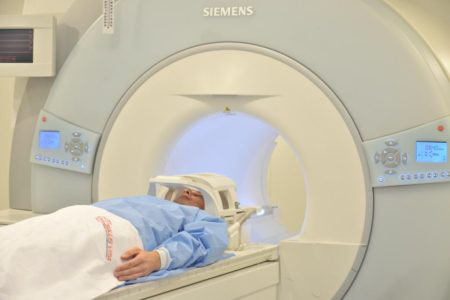
Specialist: in our clinic “SALUS-VITA” we use a 1.5 Tesla expert-class MRI scanner from SIEMENS “Espree”, which has a huge potential of software packages.
The most powerful equipment makes it possible to make a diagnosis even in the early stages of diseases.
We use smart technology; among our colleagues who work in government agencies and especially private medical centers, unfortunately, there are also specialists who use equipment less than 10?% of its capabilities, although they could use all 100. We have to not only comply with the canons, but also constantly learn and improve in order to remain professionals in our field.
Question: Working hours of the MRI department?
Specialist: at the SALUS-VITA clinic, MRI equipment operates 24 hours a day, seven days a week, and, accordingly, medical personnel who work on MRI. This creates convenience for our citizens of the Republic of Uzbekistan and everyone who needs this type of examination.
Question: Do I need any preparation for the examination?
Specialist: The most necessary condition is the absence of generally accepted contraindications to MRI examination (presence of an artificial heart pacemaker, inner ear implants, clips in the arteries of the brain, etc.). It is also important to pre-register by phone or online, where the patient is first explained and informed of what preparation is needed depending on the anatomical area being studied. Of course, an essential condition for contrast is the presence of urea and creatinine tests before contrast-enhanced MRI studies. So, since the administration of contrast in patients with renal clearance of less than 20 ml can lead to the development of nephrogenic systemic fibrosis, at least in smaller percentages, however, in the regulation “Regulations in the UK and the European Society for Pharmacological Safety and Recommendations” of 2007, where the contraindication is hypersensitivity to the components of the drug, sickle cell anemia, pregnancy period up to 2 years and clearance creatinine <20 мл/мин. Данный анализ является страховкой от нежелательных последствий препарата и самое главное служит во благо пациента.
Question: what types of contrast are performed at your SALUS-VITA clinic?
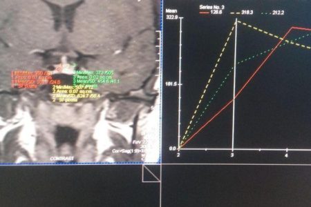
Specialist: depending on the pathology, dynamic and delayed contrast enhancement is used. In dynamic we obtain many MR sections in one anatomical area, that is, a “slice without a cut” over a certain period of time, obtaining a peak contrast curve for pathologies of the prostate, pathologies of the mammary gland, pelvic organs in women, etc. Delayed contrasts are used in demyelinating, space-occupying formations of the brain, etc. We use 0.5 molar contrast agents very widely.
For better visualization of organs and tissues, I would like to use 1.0 one-molar, organ-specific contrast agents in daily practice; unfortunately, the availability of these drugs leaves much to be desired.
Before administration After administration
Taking into account and accepting the experience of colleagues from near and far abroad, it is necessary to widely introduce into practice the use of 1.0 molar (for example: Gadovist) and organ-specific (for example: hepatotropic - Primovist) contrast agents in order to achieve the best research results in all clinics of Uzbekistan. This would make it possible to raise the scope of diagnostics an order of magnitude higher, achieving better contrast quality with a smaller injection volume.
Before administration After administration
Question: What should a doctor do if he received too much information from a study?
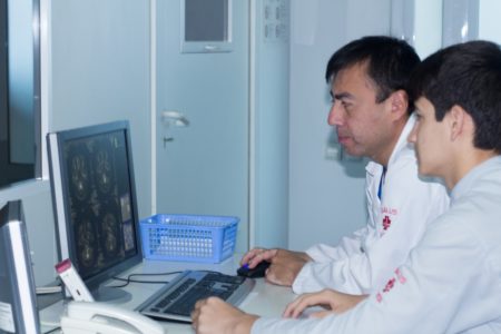
Specialist: Is there a good expression? “A radiologist sees what he knows.” If he is poorly prepared theoretically and encounters something, he will write “education, focal lesion,” which is translated into ordinary language as “I don’t know what.” And if he read and studied, he will have a differential diagnostic series in his head, that is, several diseases that may have this appearance will immediately come to mind. And he will ask the clinician to choose between them, and will suggest a way, other methods, how this can be clarified.
This is exactly what is reflected in our recommendations (recommendations of doctors at the SALUS-VITA clinic). If the image allows for several interpretations, it is necessary to talk with the patient, find out what worries him, what complaints he has, whether other examination methods were used, etc.? We often come across the opinion that the doctor is the one who guides the patient, and radiologists “only look at pictures.” And it’s okay if ordinary people think so, but sometimes clinicians are also mistaken: they think that our method is so accurate that when we receive an image, it is almost written in black and white what exactly is wrong with the patient.
Question: how to get the most out of the available information?
Specialist: Fortunately for the doctor and the patient, medicine is mostly routine, and you don’t have to solve riddles like Dr. House’s every day. But the polymorphism of pictures, pathological conditions during treatment with hormonal drugs, the state of immunodeficiency, etc. require scrupulous analysis and interpretation.
At the institute, my mentor said: a doctor should hold a picture in one hand, and “anamnesis and clinic” in the other. These rare cases are where the doctor’s qualifications are demonstrated. And how much time it took to use all the necessary program sequences in order to bring the “case” to its logical conclusion.
Let me give you an example: If you have a patient with a picture of a demyelinating disease, adhering to the McDonald diagnostic criteria of 2010, you must first exclude multiple sclerosis; for this you need to perform a contrast-enhanced MRI with a paramagnetic agent, waiting 20-30 minutes after the administration of contrast to identify this pathology. Since MS plaques tend to slowly accumulate contrast, moreover, contrast depends on the dose of administered contrast, that is, it is “time dependent” and “dose dependent”. With increasing dose of the administered contrast agent, the number of “active plaques” of multiple sclerosis increases.
At the SALUS-VITA clinic, the emphasis is on quality and individual approach to each patient!
Question: Is there a professional relationship with other specialists in clinical departments?
Specialist: In fact, in many respects, it is thanks to the work of radiologists that it is possible to make a diagnosis, and in this sense we are even in a more advantageous position than “therapists”, because we can evaluate the symptoms in the same way as they can, but we can evaluate the “pictures” much better, this is what we are “sharpened” on.
I think it is necessary to establish interdisciplinary interaction, which allows us to extract maximum benefit for the patient. For any normal radiologist, the task of “doing an MRI of the brain” is very general, it’s like saying “assembling a passenger car,” but it can be a “Tico”, or it can be a “Captiva or Orlando” with bells and whistles, and the difference between them is huge. So, it would be better for the attending physician not just to prescribe an MRI to the patient, but to set a specific task for us, radiologists and radiologists, to formulate exactly what question the study is intended to answer, so that we can carry it out as efficiently as possible.
Question: What is “terra incognita” for the diagnostics sector so far?
Specialist: Diagnostic capabilities have become much wider. For example, take adrenal gland formations, here MRI is called a non-invasive biopsy: we can tell whether it is cancer or not in almost 100?% of cases, and for us this is routine.
Molecular diagnostics are now being developed, for example, we have a contrast agent for MRI, the molecules of which are “captured” only in normal liver cells, and as a result mark them, making it possible to distinguish a good process from an evil one in the image. The buzzword “radiogenomics” has come into use?—?when a research method is used to find features in a tumor that could be somehow related to its genetics?—?after all, tumors are often caused by mutations, and we need to understand how these mutations manifest themselves macroscopically, that is, in their appearance. Is the progress good enough?—entire sets of features are being identified that can be correlated with a particular tumor genotype, and this opens up fantastic prospects.